Friday, June 29, 2012

Professor David Cooper Honored at CVPR Conference

David Cooper, Professor Emeritus of Engineering and Professor of Engineering (Research), was honored at the 25th International IEEE (Institute of Electrical and Electronics Engineers) Conference on Computer Vision and Pattern Recognition (CVPR) which was held in Providence from June 18-20. This is the major annual meeting on CVPR. Professor Cooper was honored “In appreciation of his outstanding and pioneering contributions to Unsupervised Learning and Bayesian Inference in Computer Vision.” The international conference was held this year at the Convention Center with over 1800 attendees. Brown University Professor Benjamin Kimia served as one of three general co-chairs of the conference.
Ben Kimia, David Cooper, Rama Chellappa

Professor Cooper’s current research focuses on the development and application of new geometric, algebraic, and probabilistic approaches, models, and algorithms for recognizing and estimating 2D and 3D geometric information and functioning in 3D scenes from images, video, and range data.

Professor Cooper received both his Sc.B. and Sc.M. degrees from MIT in electrical engineering, and his Ph.D. from Columbia University in applied mathematics. After graduation, he joined the Brown faculty in September of 1966 as an assistant professor. He became an associate professor in 1969, and was promoted to full professor in 1978. During his more than 45 years at Brown, Cooper has also served as cofounder and associate director of the Laboratory for Engineering Man/Machine Systems (LEMS) for more than 15 years, and the head of electrical engineering for two years. He is a fellow of the IEEE and has published roughly 140 papers in refereed journals or as book chapters.

For more information on the CVPR awards, please go to: http://www.cvpr2012.org/program-details/awards

Monday, June 25, 2012

Fei Guo Ph.D. ’12 Receives Brian Kelly Award

Brown University School of Engineering postdoctoral researcher Fei Guo Ph.D. ’12 was presented the Brian Kelly Award at Carbon 2012, the annual world conference on carbon in Krakow, Poland, on June 21. Guo, who was advised by Professor Bob Hurt at Brown, delivered a 30-minute award lecture, “Graphene-Based Environmental Barriers,” to the conference participants.

In his presentation, Guo demonstrated the potential for graphene oxide films to act as high-performance barriers for environmental toxicants. Applying elemental mercury (considered a neurotoxic) as a model, he showed that just 20 nm graphene oxide films, which were deposited onto surface treated polymers, reduced mercury permeability by 90%.

This prestigious annual award was established in 1996 by the British Carbon Group in memory of Brian Kelly, a leading authority on the physics of graphite to reward excellence in carbon science and technology. The award is currently five hundred pounds sterling (£500) and was presented at the time of the conference with a certificate. The award is intended as a travel grant for students and early career researchers with up to ten years postdoctoral experience to attend the annual World Carbon Conference.

Thursday, June 21, 2012

Selenium controls staph on implant material

A coating of selenium nanoparticles significantly reduces the growth of Staphylococcus aureus on polycarbonate, a material common in implanted devices such as catheters and endotracheal tubes, engineers at Brown University report in a new study.

PROVIDENCE, R.I. [Brown University] — Selenium is an inexpensive element that naturally belongs in the body. It is also known to combat bacteria. Still, it had not been tried as an antibiotic coating on a medical device material. In a new study, Brown University engineers report that when they used selenium nanoparticles to coat polycarbonate, the material of catheters and endotracheal tubes, the results were significant reductions in cultured populations of Staphylococcus aureus bacteria, sometimes by as much as 90 percent.

Selenium solutionQi Wang swirls a solution of selenium nanoparticles in the lab.
Coatings of the nanoparticles appear effective in fighting staph
bacteria in medical device materials, according to a new study.

Credit: Webster Lab/Brown University
“We want to keep the bacteria from generating a biofilm,” said Thomas Webster, professor of engineering and orthopaedics, who studies how nanotechnology can improve medical implants. He is the senior author of the paper, published online this week in the Journal of Biomedical Materials Research A.

Biofilms are notoriously tough colonies of bacteria to treat because they are often able to resist antibiotic drugs.

“The longer we can delay or inhibit completely the formation of these colonies, the more likely your immune system will clear them,” Webster said. “Putting selenium on there could buy more time to keep an endotracheal tube in a patient.”

Meanwhile, Webster said, because selenium is actually a recommended nutrient, it should be harmless in the body at the concentrations found in the coatings. Also, it is much less expensive than silver, a less biocompatible material that is the current state of the art for antibacterial medical device coatings.

Webster has been investigating selenium nanoparticles for years, mostly for their possible anticancer effects. As he began to look at their antibiotic properties, he consulted with Hasbro Children’s Hospital pediatrician Keiko Tarquinio, assistant professor of pediatrics, who has been eager to find ways to reduce biofilms on implants.

Studying selenium

For this study, Webster and first author Qi Wang grew selenium nanoparticles of two different size ranges and then used solutions of them to coat pieces of polycarbonate using a quick, simple process. On some of the polycarbonate, they then applied and ripped off tape not only to test the durability of the coatings but also to see how a degraded concentration of selenium would perform against bacteria.

On coated polycarbonate — both the originally coated and the tape-tested pieces — Wang and Webster used electron and atomic force microscopes to measure the concentration of nanoparticles and how much surface area of selenium was exposed to interact with bacteria.

One of their findings was that after the tape test, smaller nanoparticles adhered better to the polycarbonate than larger ones.

Then they were ready for the key step: experiments that exposed cultured staph bacteria to polycarbonate pieces, some of which were left uncoated as controls. Among the coated pieces, some had the larger nanoparticles and some had the smaller ones. Some from each of those groups had been degraded by the tape, and others had not.

All four types of selenium coatings proved effective in reducing staph populations after 24, 48, and 72 hours compared to the uncoated controls. The most potent effects — reductions larger than 90 percent after 24 hours and as much as 85 percent after 72 hours — came from coatings of either particle size range that had not been degraded by the tape. Among those coatings that had been subjected to the tape test, the smaller nanoparticle coatings proved more effective.

Staph populations exposed to any of the coated polycarbonate pieces peaked at the 48-hour timeframe, perhaps because that is when the bacteria could take fullest advantage of the in vitro culture medium. But levels always fell back dramatically by 72 hours.

The next step, Webster said, is to begin testing in animals. Such in vivo experiments, he said, will test the selenium coatings in a context where the bacteria have more available food but will also face an immune system response.

The results may ultimately have commercial relevance. Former graduate students developed a business plan for the selenium nanoparticle coatings while in school and have since licensed the technology from Brown for their company, Axena Technologies.

Monday, June 11, 2012

Small Wonder

Partnering with an engineer, a pathologist goes in a new direction.

The yellow-and-black signs outside Dr. Agnes Kane’s pathology laboratory read “CAUTION: Cancer hazard.” Nodding at the ominous-looking postings, Kane explains, “because of their toxicity similar to asbestos, we handle these materials as if they were carcinogens.” Meanwhile, across the Providence River, at the School of Engineering, Professor Robert Hurt is hard at work creating the very materials that Kane is so gingerly studying: nanoparticles.

Smaller than 1,000th the width of a human hair—so small that you need an electron microscope to see them— nanoparticles’ practical applications may be enormous: making implants more biocompatible; diagnosing and treating cancers; cleaning up oil spills. That said, the history of science is filled with promising solutions that create additional unforeseen problems of their own. No one is more aware of this than Kane, chair of Brown’s Department of Pathology and Laboratory Medicine. She has spent her career on, and helped guide the Department’s focus on, the human health effects of environmental and occupational exposures. She and Hurt tick off some examples demonstrating this law of unintended consequences:

“Corn ethanol,” says Hurt, referring to the fact that 40 percent of the corn grown in America is used to create this alternative fuel. “Then you raise the corn prices for food.”

Kane nods. “Use more fertilizer? Contaminate our water supplies. There’s always these trade-offs.”

One of modern history’s most devastating trade-offs was of a common mineral that makes an excellent flameretardant building material. Its usefulness notwithstanding, asbestos can cause devastating cancers and fatal lung problems both for those who mine it and for those who live and work in buildings that contain it.

Small, Novel...but Safe

Selenium-carbon nanocomposite particles
synthesized as a novel chemotherapy agent

From the time Kane joined Brown’s pathology department as a founding member in 1982, she has studied the mechanisms by which asbestos injures cells and causes cancer. When, in 2004, she gave a talk about this research to a group of colleagues, Hurt approached her afterward. The asbestos fibers that Kane showed in her talk reminded Hurt of the carbon nanofibers he had been developing. “We were not working on health effects at the time,” Hurt says. “We were doing traditional nanoscience, trying to make new things that had never been made before.”

But when Hurt told Kane about his carbon nanofibers, “I immediately asked him if I could have some,” Kane recalls. Her worrisome discovery—that the particles were similar to asbestos in several key ways—has changed the direction of both her own and Hurt’s careers and of the pathology department’s research and teaching.

Now Kane and Hunt work side-by-side to create innovative nanotechnology and, simultaneously, assess the materials’ safety and toxicity. “It’s a new paradigm to try to consider the implications of the technology as you develop the technology,” says Hurt. “We haven’t done a lot of that in the past. We just develop technology and we field it and then we worry about what its implications might be. So it’s kind of fun to do these things together.”

In 2007, their collaboration gave rise to the Institute for Molecular and Nanoscale Innovation (IMNI), an interdisciplinary organization comprising more than 60 faculty in nine departments. Kane heads IMNI’s NanoHealth Initiative, which studies the environmental and health effects of nanotechnology.

Training the Next Interdisciplinarians

With curly chin-length gray hair and blue eyes, Kane—known to friends and colleagues as “Aggie”—smiles often and laughs readily. Her unassuming manner and commitment to collaboration, teaching, and mentorship have won her numerous teaching awards and devotees.

“If it weren’t for Aggie, I wouldn’t be doing what I’m doing,” says Luba Dumenco, a lecturer in pathology and director of the Medical School’s preclinical curriculum. “She’s always valued teaching incredibly highly.” Just recently, Dumenco struck up a conversation with another mom at the local skating rink.The woman happened to be a neonatologist who had trained at Brown’s medical school. “I told her I was teaching at the med school, and she said, ‘Do you know Dr. Aggie Kane? She was our favorite! We loved her!’” Dumenco says with a laugh. “She cares a lot about the students.She does a wonderful job and they’re very lucky to have her.”

The breadth of students that Kane reaches each year has grown as a result of her partnership with Hurt. In 2009, they secured a grant from GAANN, or Graduate Assistance in Areas of National Need, to fund interdisciplinary training in nanotechnology. Between six and eight doctoral students study nanotoxicology and nanomedicine with co-mentors in engineering or physical sciences and biological science. Kane and Hurt also co-teach an undergraduate and graduate course called “Small Wonders: Science, Technology, and Human Health Impacts of Nanomaterials.” For their final projects, students working together in interdisciplinary teams are required both to use nanotechnology to solve some real-world problem and to address—and minimize—their solution’s potential environmental and health impacts. “I look at this as training the next generation of environmental scientists and engineers,” Kane says.

But first they have to learn how to talk to each other. When Kane and Hurt began collaborating, “it took us a while to learn each other’s languages,” says Kane, “because medicine has its own vocabulary, as well as engineering.” Kane might, for example, say “mitochondria,” or “epigenetics,” and get a blank stare in return. “And so we would just keep asking each other questions, any time we didn’t understand something,” she recalls. “It took us quite some time to learn enough to communicate effectively.”

Their newest collaboration is funded by the Gulf of Mexico Research Initiative, which was established in the wake of the Deepwater Horizon disaster. Hurt has set out to design nanoparticles called nanosorbents, which by capturing and sequestering pollutants like oil, may be safer and more effective than existing methods of cleaning up oil spills. The Deepwater Horizon cleanup team—like the Exxon Valdez team before it—relied on Corexit, a dispersant which causes oil to suspend in the water as tiny particles rather than accumulate on the surface as oil slicks.

“They used it in enormous amounts in the Deepwater Horizon cleanup,” says Hurt, but “it’s not clear if it’s a good idea to use very large amounts of chemicals in a marine environment.”

But it’s not clear whether nanosorbents are a good idea, either. As Hurt designs the particles, Kane and her team set out to answer two questions. “First, will they work?” she asks. “And then, will they be toxic to the organisms?”

“They might be worse,” Hurt acknowledges. “We don’t know.”

Engineering Prevention

To begin to answer these questions, Kane has a small steel tank in her lab. Like a miniature wave pool, the open-air tank bubbles with seawater maintained at exactly 72 degrees. Soon this will be home to a small colony of brine shrimp, tiny marine organisms that, as larvae in the wild, are eaten by small fish, which, in turn, are used as bait to catch larger fish, which are eaten by people. As such, the brine shrimp are a good “indicator species” for study.

“We don’t want to have these kinds of dispersants accumulate up the food chain,” says Kane, peeking at the churning water.

A tube runs from a beaker into the basin, helping to aerate the water. As the shrimp grow in the lab, Kane and her colleagues will release oil and Hurt’s nanoparticles into the water with them to see what happens. Will they stop swimming? Will they die? Will their RNA reflect toxicity or injury? If so, Kane says, she is confident that her colleagues can alter the nanoparticles to reflect her findings.

“Engineers are very clever,” she says with a smile. “If we can identify the specific properties that are associated with the toxic effects, they can design [the nanoparticles] or process them to eliminate those properties or reduce those properties and reduce their toxicity.” And part of the excitement of studying nanoparticles is the ability to intervene now, in the very early stages—to prevent environmental and health disasters, rather than clean them up after the fact.

“When you think about what happened with the widespread use of asbestos throughout the 20th century— and we’re still suffering the consequences because of the long latent period of those diseases—the fact that those fibers persist in the buildings and in the environment and we’re still being exposed,” says Kane, “that’s a very expensive lesson. We do not want to repeat that tragedy again.”

by Beth Schwartzapfel ’01
Photographs by Karen Philippi
Courtesy of Brown Medicine Magazine

A SMART(er) way to track influenza

Brown University researchers have created a reliable and fast flu-detection test that can be carried in a first-aid kit. The novel prototype device isolates influenza RNA using a combination of magnetics and microfluidics, then amplifies and detects probes bound to the RNA. The technology could lead to real-time tracking of influenza. Results are published in the Journal of Molecular Diagnostics.

PROVIDENCE, R.I. [Brown University] — In April 2009, the world took notice as reports surfaced of a virus in Mexico that had mutated from pigs and was being passed from human to human. The H1N1 “swine flu,” as the virus was named, circulated worldwide, killing more than 18,000 people, according to the World Health Organization. The Centers for Disease Control and Prevention in the United States said it was the first global pandemic in more than four decades.

Swine flu will not be the last viral mutation to cause a worldwide stir. One way to contain the next outbreak is by administering tests at the infection’s source, pinpointing and tracking the pathogen’s spread in real time. But such efforts have been stymied by devices that are costly, unwieldy and unreliable. Now, biomedical engineers at Brown University and Memorial Hospital in Rhode Island have developed a biochip that can detect the presence of influenza by zeroing in on the specific RNA sequence and then using tiny magnets in a tube to separate the flu-ridden sequence from the rest of the RNA strand. The result: A reliable, fast prototype of a flu-detection test that potentially can be carried in a first-aid kit and used as easily as an iPhone.

“We wanted to make something simple,” said Anubhav Tripathi, associate professor of engineering at Brown and the corresponding author on the paper, published in the Journal of Molecular Diagnostics. “It’s a low-cost device for active, on-site detection, whether it’s influenza, HIV, or TB (tuberculosis).”

The Brown assay is called SMART, which stands for “A Simple Method for Amplifying RNA Targets.” Physically, it is essentially a series of tubes, with bulbs on the ends of each, etched like channels into the biochip.

There are other pathogen-diagnostic detectors, notably the Polymerase Chain Reaction device (which targets DNA) and the Nucleic Acid Sequence Based Amplification (which also targets RNA). The SMART detector is unique in that the engineers use a DNA probe with base letters that match the code in the targeted sequence. This ensures the probe will latch on only to the specific RNA strand being assayed. The team inundates the sample with probes, to ensure that all RNA molecules bind to a probe.

“The device allows us to design probes that are both sensitive and specific," Tripathi said.


Anubhav Tripathi
“We wanted to make something simple. (This is) a low-cost device
for active, on-site detection, whether it’s influenza, HIV, or TB.”

Credit: Mike Cohea/Brown University
This approach creates excess — that is, probes with no RNA partners. That’s OK, because the Brown-led team then attached the probes to 2.8 micron magnetic beads that carry the genetic sequence for the influenza RNA sequence. The engineers then use a magnet to slowly drag the RNA-probe pairs collected in the bulb through a tube that narrows to 50 microns and then deposit the probes at a bulb at the other end. This convergence of magnetism (the magnetized probes and the dragging magnets) and microfluidics (the probes’ movement through the narrowing channel and the bulbs) serves to separate the RNA-probe pairs from the surrounding biological debris, allowing clinicians to isolate the influenza strains readily and rapidly for analysis. The team reports that it tracks the RNA-probe beads flawlessly at speeds up to 0.75 millimeters per second.

“When we amplify the probes, we have disease detection,” Tripathi said. “If there is no influenza, there will be no probes (at the end bulb). This separation part is crucial.”

Once separated, or amplified, the RNA can be analyzed using conventional techniques, such as nucleic acid sequence-based amplification (NASBA).

The chips created in Tripathi’s lab are less than two inches across and can fit four tube-and-bulb channels. Tripathi said the chips could be commercially manufactured and made so more channels could be etched on each.

The team is working on separate technologies for biohazard detection.

Stephanie McCalla, who earned her doctorate at Brown last year and is now at the California Institute of Technology, is the first author on the paper. Brown professors of medicine Steven Opal and Andrew Artenstein, with Carmichael Ong and Aartik Sarma, who earned their undergraduate degrees at Brown, are contributing authors.

The U.S. National Institutes of Health and the National Science Foundation funded the research.

- by David Orenstein

Wednesday, May 23, 2012

How ion bombardment reshapes metal surfaces

Ion bombardment of metal surfaces is an important, but poorly understood, nanomanufacturing technique. New research using sophisticated supercomputer simulations has shown what goes on in trillionths of a second. The advance could lead to better ways to predict the phenomenon and more uses of the technique to make new nanoscale products.

PROVIDENCE, R.I. [Brown University] — To modify a metal surface at the scale of atoms and molecules — for instance to refine the wiring in computer chips or the reflective silver in optical components — manufacturers shower it with ions. While the process may seem high-tech and precise, the technique has been limited by the lack of understanding of the underlying physics. In a new study, Brown University engineers modeled noble gas ion bombardments with unprecedented richness, providing long-sought insights into how it works.

Three new mechanisms at the nanoscale
A computer-model image of an island of metal atoms
formed after bombardment by noble gas ions. Atoms
disturbed by the bombardment cluster together under
the surface and then glide back up in a matter of 2.1
trillionths of a second, or picoseconds (ps).

Credit: Kim Lab/Brown University
“Surface patterns and stresses caused by ion beam bombardments have been extensively studied experimentally but could not be predicted accurately so far,” said Kyung-Suk Kim, professor of engineering at Brown and co-author of the study published May 23 in the Proceedings of the Royal Society A. “The new discovery is expected to provide predictive design capability for controlling the surface patterns and stresses in nanotechnology products.”

The improved understanding could open the door to new technologies, Kim said, such as new approaches to make flexible electronics, biocompatible surfaces for medical devices, and more damage-tolerant and radiation-resistant surfaces. The research applies to so-called “FCC” metals such as copper, silver, gold, nickel, and aluminum. Those metals are crystals made up of cubic arrangements of atoms with one at each corner and one in each cube-face center.

Scientists have been trying to explain the complicated process for decades, and more recently they have begun to try modeling it on computers. Kim said the analysis of the Brown team, including lead author and postdoctoral scholar Sang-Pil Kim, was more sophisticated than previous attempts that focused on a single bombardment event and only isolated point defects within the metal substrate.

“In this work, for the first time, we investigate collective behavior of those defects during ion bombardments in terms of ion-substrate combinations,” Kyung-Suk Kim said.

The new model revealed how ion bombardments can set three main mechanisms into motion in a matter of trillionths of a second. The researchers dubbed the mechanisms “dual layer formation,” “subway-glide mode growth,” and “adatom island eruption.” They are a consequence of how the incoming ions melt the metal and then how it resolidifies with the ions occasionally trapped inside.

When ions hit the metal surface, they penetrate it, knocking away nearby atoms like billiard balls in a process that is akin, at the atomic level, to melting. But rather than merely rolling away, the atoms are more like magnetic billiard balls in that they come back together, or resolidify, albeit in a different order.

Some atoms have been shifted out of place. There are some vacancies in the crystal nearer to the surface, and the atoms there pull together across the empty space, that creates a layer with more tension. Beneath that is a layer with more atoms that have been knocked into it. That crowding of atoms creates compression. Hence there are now two layers with different levels of compression and tension.This “dual layer formation” is the precursor to the “subway-glide mode growth” and “adatom island eruption”.

A hallmark of materials that have been bombarded with ions is that they sometimes produce a pattern of material that seems to have popped up out of the original surface. Previously, Kyung-Suk Kim said, scientists thought displaced atoms would individually just bob back up to the surface like fish killed in an underwater explosion. But what the team’s models show is that these molecular islands are formed by whole clusters of displaced atoms that bond together and appear to glide back up to the surface.

“The process is analogous to people getting on a subway train at suburban stations, and they all come out together to the surface once the train arrives at a downtown station during the morning rush hour,” Kyung-Suk Kim said.

The mechanisms, while offering a new explanation for the effects of ion bombardment, are just the beginning of this research.

 “As a next step, I will develop prediction models for nanopattern evolution during ion bombardment which can guide the nanomanufacturing processes,” Sang-Pil Kim said. “This research will also be expanded to other applications such as soft- or hard-materials under extreme conditions.”

In addition to Kyung-Suk Kim and Sang-Pil Kim, other authors include Huck Beng Chew, Eric Chason and Vivek Shenoy.

The research was funded by the Korea Institute of Science and Technology, the U.S. National Science Foundation, and the U.S. Department of Energy. The work used the Extreme Science and Engineering Discovery Environment (XSEDE), which is supported by National Science Foundation grant number OCI-1053575.

Wednesday, May 16, 2012

People with paralysis control robotic arms using brain-computer interface

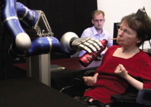
A new study in Nature reports that two people with tetraplegia were able to reach for and grasp objects in three-dimensional space using robotic arms that they controlled directly with brain activity. They used the BrainGate neural interface system, an investigational device currently being studied under an Investigational Device Exemption. One participant used the system to serve herself coffee for the first time since becoming paralyzed nearly 15 years ago.
One small step  
A 58-year-old woman, paralyzed by a stroke for almost 15 years, uses her thoughts to control a robotic arm, grasp a bottle of coffee, serve herself a drink, and return the bottle to the table.

PROVIDENCE, R.I. [Brown University] — On April 12, 2011, nearly 15 years after she became paralyzed and unable to speak, a woman controlled a robotic arm by thinking about moving her arm and hand to lift a bottle of coffee to her mouth and take a drink. That achievement is one of the advances in brain-computer interfaces, restorative neurotechnology, and assistive robot technology described in the May 17 edition of the journal Nature by the BrainGate2 collaboration of researchers at the Department of Veterans Affairs, Brown University, Massachusetts General Hospital, Harvard Medical School, and the German Aerospace Center (DLR).

A 58-year-old woman (“S3”) and a 66-year-old man (“T2”) participated in the study. They had each been paralyzed by a brainstem stroke years earlier which left them with no functional control of their limbs. In the research, the participants used neural activity to directly control two different robotic arms, one developed by the DLR Institute of Robotics and Mechatronics and the other by DEKA Research and Development Corp., to perform reaching and grasping tasks across a broad three-dimensional space. The BrainGate2 pilot clinical trial employs the investigational BrainGate system initially developed at Brown University, in which a baby aspirin-sized device with a grid of 96 tiny electrodes is implanted in the motor cortex — a part of the brain that is involved in voluntary movement. The electrodes are close enough to individual neurons to record the neural activity associated with intended movement. An external computer translates the pattern of impulses across a population of neurons into commands to operate assistive devices, such as the DLR and DEKA robot arms used in the study now reported in Nature.

BrainGate participants have previously demonstrated neurally based two-dimensional point-and-click control of a cursor on a computer screen and rudimentary control of simple robotic devices.

The study represents the first demonstration and the first peer-reviewed report of people with tetraplegia using brain signals to control a robotic arm in three-dimensional space to complete a task usually performed by their arm. Specifically, S3 and T2 controlled the arms to reach for and grasp foam targets that were placed in front of them using flexible supports. In addition, S3 used the DLR robot to pick up a bottle of coffee, bring it to her mouth, issue a command to tip it, drink through a straw, and return the bottle to the table. Her BrainGate-enabled, robotic-arm control during the drinking task required a combination of two-dimensional movements across a table top plus a “grasp” command to either grasp and lift or tilt the robotic hand.

“Our goal in this research is to develop technology that will restore independence and mobility for people with paralysis or limb loss,” said lead author Dr. Leigh Hochberg, a neuroengineer and critical care neurologist who holds appointments at the Department of Veterans Affairs, Brown University, Massachusetts General Hospital, and Harvard. He is the sponsor-investigator for the BrainGate2 pilot clinical trial. “We have much more work to do, but the encouraging progress of this research is demonstrated not only in the reach-and-grasp data, but even more so in S3’s smile when she served herself coffee of her own volition for the first time in almost 15 years.”
Leigh HochbergEven after nearly 15 years, a part of the brain essentially “disconnected” from its original target by a brainstem stroke was still able to direct the complex, multidimensional movement of an external arm.
Leigh Hochberg
Even after nearly 15 years, a part of the brain essentially “disconnected” from its
original target by a brainstem stroke was still able to direct the complex,
multidimensional movement of an external arm.
Partial funding for this work comes from the VA, which is committed to improving the lives of injured veterans. “VA is honored to have played a role in this exciting and promising area of research,” said VA Secretary Eric Shinseki. “Today’s announcement represents a great step forward toward improving the quality of life for veterans and others who have either lost limbs or are paralyzed.”

Hochberg adds that even after nearly 15 years, a part of the brain essentially “disconnected” from its original target by a brainstem stroke was still able to direct the complex, multidimensional movement of an external arm — in this case, a robotic limb. The researchers also noted that S3 was able to perform the tasks more than five years after the investigational BrainGate electrode array was implanted. This sets a new benchmark for how long implanted brain-computer interface electrodes have remained viable and provided useful command signals.

John Donoghue, the VA and Brown neuroscientist who pioneered BrainGate more than a decade ago and who is co-senior author of the study, said the paper shows how far the field of brain-computer interfaces has come since the first demonstrations of computer control with BrainGate.

“This paper reports an important advance by rigorously demonstrating in more than one participant that precise three-dimensional neural control of robot arms is not only possible, but also repeatable,” said Donoghue, who directs the Brown Institute for Brain Science. “We’ve moved significantly closer to returning everyday functions, like serving yourself a sip of coffee, usually performed effortlessly by the arm and hand, for people who are unable to move their own limbs. We are also encouraged to see useful control more than five years after implant of the BrainGate array in one of our participants. This work is a critical step toward realizing the long-term goal of creating a neurotechnology that will restore movement, control, and independence to people with paralysis or limb loss.”

In the research, the robots acted as a substitute for each participant’s paralyzed arm. The robotic arms responded to the participants’ intent to move as they imagined reaching for each foam target. The robot hand grasped the target when the participants imagined a hand squeeze. Because the diameter of the targets was more than half the width of the robot hand openings, the task required the participants to exert precise control. (Videos of these actions are available on the Nature website.)
John Donoghue“We’ve moved significantly closer to returning everyday functions, like serving yourself a sip of coffee, usually performed effortlessly by the arm and hand, for people who are unable to move their own limbs.”
John Donoghue
“We’ve moved significantly closer to returning everyday functions, like serving yourself
a sip of coffee, usually performed effortlessly by the arm and hand, for people who are
unable to move their own limbs.”
In 158 trials over four days, S3 was able to touch the target within an allotted time in 48.8 percent of the cases using the DLR robotic arm and hand and 69.2 percent of the cases with the DEKA arm and hand, which has the wider grasp. In 45 trials using the DEKA arm, T2 touched the target 95.6 percent of the time. Of the successful touches, S3 grasped the target 43.6 percent of the time with the DLR arm and 66.7 percent of the time with the DEKA arm. T2’s grasp succeeded 62.2 percent of the time.

T2 performed the session in this study on his fourth day of interacting with the arm; the prior three sessions were focused on system development. Using his eyes to indicate each letter, he later described his control of the arm: “I just imagined moving my own arm and the [DEKA] arm moved where I wanted it to go.”

The study used two advanced robotic arms: the DLR Light-Weight Robot III with DLR five-fingered hand and the DEKA Arm System. The DLR LWR-III, which is designed to assist in recreating actions like the human arm and hand and to interact with human users, could be valuable as an assistive robotic device for people with various disabilities. Patrick van der Smagt, head of bionics and assistive robotics at DLR, director of biomimetic robotics and machine learning labs at DLR and the Technische Universität München, and a co-senior author on the paper said: “This is what we were hoping for with this arm. We wanted to create an arm that could be used intuitively by varying forms of control. The arm is already in use by numerous research labs around the world who use its unique interaction and safety capabilities. This is a compelling demonstration of the potential utility of the arm by a person with paralysis.”

DEKA Research and Development developed the DEKA Arm System for amputees, through funding from the United States Defense Advanced Research Projects Agency (DARPA). Dean Kamen, founder of DEKA said, “One of our dreams for the Luke Arm [as the DEKA Arm System is known informally] since its inception has been to provide a limb that could be operated not only by external sensors, but also by more directly thought-driven control. We’re pleased about these results and for the continued research being done by the group at the VA, Brown and MGH.” The research is aimed at learning how the DEKA arm might be controlled directly from the brain, potentially allowing amputees to more naturally control this prosthetic limb.

Over the last two years, VA has been conducting an optimization study of the DEKA prosthetic arm at several sites, with the cooperation of veterans and active duty service members who have lost an arm. Feedback from the study is helping DEKA engineers to refine the artificial arm’s design and function. “Brain-computer interfaces, such as BrainGate, have the potential to provide an unprecedented level of functional control over prosthetic arms of the future,” said Joel Kupersmith, M.D., VA chief research and development officer. “This innovation is an example of federal collaboration at its finest.”
The BrainGate2 Neural Interface SystemAn implanted microelectrode array, first used more than a decade ago, detects brain signals which can be translated by a computer into machine instructions, allowing control of robotic devices by thought.
The BrainGate2 Neural Interface System
An implanted microelectrode array, first used more than a decade ago,
detects brain signals which can be translated by a computer into machine
instructions, allowing control of robotic devices by thought.

Story Landis, director of the National Institute of Neurological Disorders and Stroke, which funded the work in part, noted: “This technology was made possible by decades of investment and research into how the brain controls movement. It’s been thrilling to see the technology evolve from studies of basic neurophysiology and move into clinical trials, where it is showing significant promise for people with brain injuries and disorders.”

In addition to Hochberg, Donoghue, and van der Smagt, other authors on the paper are Daniel Bacher, Beata Jarosiewicz, Nicolas Masse, John Simeral, Joern Vogel, Sami Haddadin, Jie Liu, and Sydney Cash.

Additional comments

Vincent Ng
Medical Center Director, Providence VA Medical Center
“The VA is on the forefront of translational research that’s improving the quality of life for our Veterans who have sacrificed so much for our Nation. We are proud to be a part of this exciting, collaborative research.”

U.S. Sen. Sheldon Whitehouse

“I congratulate Brown University and the Providence VA Medical Center for this ground-breaking project, which could help to significantly improve the quality of life of disabled and paralyzed Americans, including many veterans. The innovations produced in this new study highlight the value of federal support for basic scientific research.”

U.S. Rep. David Cicilline

“I congratulate the entire Brown University community on the progress it has made in this project. It is my hope that with continued success, this advancement will help improve the quality of life for individuals with disabilities, especially our men and women in uniform.”

Jennifer French

Executive Director, Neurotech Network
“This latest development in cortical control research has the potential to revolutionize the way we interact with technology. More specifically, the possibilities open a new level of independence for those living with severe paralysis. Simple tasks like drinking, eating or brushing your teeth are not possible for people living with severe paralysis. The ability to perform these every-day tasks can create a new world of independence for people with severe disabilities.”

R. John Davenport

Associate Director, Brown University Institute for Brain Science
“This exciting advance from the BrainGate team exemplifies the amazing science that can only result when researchers from disparate disciplines collaborate. The Institute works to link fundamental science, engineering, and medicine among our more than 100 faculty members.”
The BrainGate2 study continues to enroll participants to take part in this research and recently added Stanford University as a member of the collaboration and a clinical trial site.

About the BrainGate collaboration

This advance is the result of the ongoing collaborative BrainGate research at Brown University, Massachusetts General Hospital, Providence VA Medical Center; researchers at Stanford University have recently joined the collaboration as well. The BrainGate research team is focused on developing and testing neuroscientifically inspired technologies to improve the communication, mobility, and independence of people with neurologic disorders, injury, or limb loss.

Funding for the study and its projects comes from the Rehabilitation Research and Development Service, Office of Research and Development, U.S. Department of Veterans Affairs, the National Institutes of Health (some grants were funded all or in part through the American Recovery and Reinvestment Act), the Eunice Kennedy Shriver National Institute of Child Health and Human Development/National Center for Medical Rehabilitation Research (HD53403, HD100018, HD063931), the National Institute on Deafness and Other Communication Disorders, the National Institute of Neurological Disorders and Stroke (NS025074), the National Institute of Biomedical Imaging and Bioengineering (EB007401), the Doris Duke Charitable Foundation, the MGH-Deane Institute for Integrated Research on Atrial Fibrillation and Stroke, Katie Samson Foundation, and the Craig H. Neilsen Foundation. The contents do not represent the official views of the Department of Veterans Affairs or the United States Government.

The implanted microelectrode array and associated neural recording hardware used in the BrainGate research are manufactured by BlackRock Microsystems LLC (Salt Lake City, Utah). The research prototype Gen2 DEKAarm was provided by DEKA Integrated Solutions Inc, under contract from the Defense Advanced Research Project Agency (DARPA).

The BrainGate pilot clinical trial was previously directed by Cyberkinetics Neurotechnology Systems Inc. Foxborough, Mass., (CKI). CKI ceased operations in 2009, before the collection of data reported in the Nature manuscript. The clinical trials of the BrainGate2 Neural Interface System are now administered by Massachusetts General Hospital, Boston, Mass. Donoghue is a former chief scientific officer and a former director of CKI; he held stocks and received compensation. Hochberg received research support from Massachusetts General and Spaulding Rehabilitation Hospitals, which in turn received clinical trial support from Cyberkinetics.

CAUTION: Investigational Device. Limited by Federal Law to Investigational Use. The device is being studied under an IDE for the detection and transmission of neural signals from the cortex to externally powered communication systems, environmental control systems, and assistive devices by persons unable to use their hands due to physical impairment. The clinical trial is ongoing; results presented are thus preliminary. The safety and effectiveness of the device have not been established.

Press contacts
David Orenstein, Brown University, david_orenstein@brown.edu, 401-527-2525
Mark Ballesteros, U.S. Dept. of Veterans Affairs, Mark.Ballesteros@va.gov, 202-461-7559.
Michael Morrison, Massachusetts General Hospital, mdmorrison@partners.org, 617-724-6425

Tuesday, May 15, 2012

Brian Reggiannini figures out who’s talking

If computers could become ‘smart’ enough to recognize who is talking, that could allow them to produce real-time transcripts of meetings, courtroom proceedings, debates, and other important events. In the dissertation that will allow him to receive his Ph.D. at Commencement this year, Brian Reggiannini found a way to advance the state of the art for voice- and speaker-recognition.

Real-time tracking of who’s talking
With the right algorithms and signal processing software, an
array of button-size microphones placed around the perimeter
of a room can identify, follow, and record each of several
people as they move about, interrupt each other, and converse.

Credit: Frank Mullin/Brown University
Everyone does signal processing every day, even if we don’t call it that. With friends at a sports bar, we peer up at the TV to see the score, we turn our head toward the crashing sound when a waitress drops a glass, and perhaps most remarkably, we can track the fast-paced banter of all the people in our booth, even if we’ve never met some of the friends-of-friends who have insinuated themselves into the scene.

Very few of us, however, could ever get a computer to do anything like that. That’s why doing it well has earned Brian Reggiannini a Ph.D. at Brown and a career in the industry.

In his dissertation, Reggiannini managed to raise the bar for how well a computer connected to a roomful of microphones can keep track of who among a small group of speakers is talking. Further refined and combined with speech recognition, such a system could lead to instantaneous transcriptions of meetings, courtroom proceedings, or debates among, say, several rude political candidates who are prone to interrupt. It could help the deaf follow conversations in real-time.

If only it weren’t so hard to do.

Brian Reggiannini“We’re trying to teach a computer how to do something that we as humans do so naturally that we don’t even understand how we do it.”Brian Reggiannini
“We’re trying to teach a computer how to do something that we as humans do so naturally that we don’t even understand how we do it.”

But Reggiannini, who came to Brown as an undergraduate in 2003 and began building microphone arrays in the lab of Harvey Silverman, professor of engineering, in his junior year, was determined to advance the state of the art.
The specific challenge he set for himself was real-time tracking of who’s talking among at least a few people who are free to rove around a room. Hardware was not the issue. The test room on campus has 448 microphones all around the walls and he only used 96. That was enough to gather the kind of information that allows systems – think of your two ears – to locate the source of a sound.
The real rub was in devising the algorithms and, more abstractly, in realizing where his reasoning about the problem had to abandon the conventional wisdom.
Previous engineers who had tried something like this were on the right track. After all, there is only so much data available in situations like this. Some tried analyzing accents, pronunciation, word use, and cadence, but those are complex to track and require a lot of data. The simpler features are pitch, volume, and spectral statistics (a breakdown of a voice’s component waves and frequencies) of each speaker’s voice. Systems can also ascertain where a voice came from within the room.
Snippets, not speakers
But many attempts to build speaker identification systems (like the voice recognition in your personal computer) have relied on the idea that a computer could be extensively trained in “clean,” quiet conditions to learn a speaker’s voice in advance.
One of Reggiannini’s key insights was that just like a politician couldn’t possibly be primed to recognize every voter at a rally, it’s unrealistic to train a speaker-recognition system with the voice of everyone who could conceivably walk into a room.
Instead, Reggiannini sought to build a system that could learn to distinguish the voices of anyone within a session. It analyzes each new segment of speech and also notes the distinct physical position of individuals within the room. The system compares each new segment, or snippet, of what it hears to previous snippets. It then determines a statistical likelihood that the new snippet would have come from a speaker it has already identified as unique.
“Instead of modeling talkers, I’m going to instead model pairs of speech segments,” Reggiannini recalled.
A key characteristic of Reggiannini’s system is that it can work with very short snippets of speech. It doesn’t need full sentences to work at least somewhat well. That’s important because it’s realistic. People don’t speak in florid monologues. They speak in fractured conversations. No way! Yes, really.
People also are known to move around. For that reason position as inferred by the array of microphones can be only an intermittent asset. At any single moment in time, especially at the beginning of a session, position helpfully distinguishes each talker from every other (no two people can be in the same place at the same time), but when people stop talking and start walking, the system necessarily loses track of them until they speak again.
Reggiannini tested his system every step of the way. His experiments included just pitch analysis, just spectral analysis, a combination of the two, position alone, and a combination of the full speech analysis and position tracking. He subjected the system to a multitude of voices, sometimes male-only, sometimes female-only, and sometimes mixed. In every case, at least until the speech snippets became quite long, his system was better able to discriminate among talkers than two other standard approaches.
That said, the system sometimes is uncertain and in cases like that it defers assigning speech to a talker until it is more certain. Once it is, it goes back and labels the snippets accordingly.
It’s no surprise that the system would err, or hedge, here and there. Reggiannini’s test room was noisy. While some systems are fed very clean audio, the only major concessions that Reggiannini allowed himself were that speakers wouldn’t run or jump across the room and that only one would speak from the script at a time. The ability to filter individual voices out from within overlapping speech is perhaps the biggest remaining barrier between the system remaining a research project and becoming a commercial success.
A career in the field
While the ultimate fate of Reggiannini’s innovations is not yet clear, what is certain is that he has been able to embark on a career in the field he loves. Since leaving Brown last summer he’s been working as a digital signal processing engineer at Analog Devices in Norwood, Mass., which happens to be his hometown.
Reggiannini has yet to work on an audio project, but that’s fine with him. His interest is the signal processing, not sound per se. Instead he’s applied his expertise to challenges of heart monitoring and wireless communications.
“I’ve been jumping around applications but all the fundamental signal processing theory applies no matter what the signal is,” he said. “My background lets me work on a wide range of problems.”
After seven years and three degrees at Brown, Reggiannini was prepared to pursue his passion.

- by David Orenstein

Friday, May 4, 2012

Brown Team Wins Rhode Island Business Plan Competition For Fourth Straight Year

For the fourth consecutive year, a Brown student team has won the Rhode Island Business Plan Competition. This year, Overhead.fm, led by Stephen Hebson ’12, an economics and history double concentrator, and Parker Wells ’12, a mechanical engineering concentrator, won the student track of the competition. Their plan is to produce a web app to provide a customizable stream of music for in-store use, licensing music directly from the artists and labels. They received $15,000 in cash and services valued at $24,000 for winning the competition. Winners were announced at the annual RIBX business expo at the Rhode Island Convention Center. In order to be eligible to win, applicants had to agree to establish or maintain operations in Rhode Island.

Previous Brown winners of the competition have included PriWater, now Premama, (2011), Speramus (2010), and Runa (2009). Premama (http://drinkpremama.com/) produces a prenatal beverage supplement to help reduce birth defects. Speramus is (www.speramus.com) an online fundraising platform that matches donors with individual support opportunities. Runa (www.runa.org) produces energy drinks made from the leaves of an Amazonian tree, and has been featured in the New York Times and raised over $1 million from investors and has increased distribution into Whole Foods.

Two other Brown teams, both with connections to the School of Engineering, were also named finalists at the 2012 competition. Finalists receive $5,000 in cash and services valued at between $9,000 and $11,500.

One of the finalists, JCD Wind, was from Steve Petteruti’s Entrepreneurship I class and Entrepreneurship II classes, Engineering 1930G and Engineering 1930H. JCD Wind, included James McGinn ’12, a biomedical engineering concentrator, and Carli Wiesenfeld ’12, a commerce, organizations, and entrepreneurship (COE) concentrator. The company aims to make seamless, high strength lightweight carbon fiber turbine blades. McGinn is also a member of the men’s rugby team, and Wiesenfeld is a member of the women’s gymnastics team.

The other finalist was a group of four graduate students in the Program in Innovation Management and Entrepreneurship (PRIME) master’s degree program. Solar4Cents included PRIME students Sean Pennino, Bhavuk Nagpal, Xiaotong 'Peter' Shan, and Meng 'Milo' Zhang. Solar4Cents is a manufacturing company that aims to produce low-cost, thin-film copper, zinc, tin, and sulfur solar cells for solar panel manufacturers.

Established in 2000, the Rhode Island Business Plan Competition has awarded more than $1 million in prizes since its inception.

For the official RI Business plan release on the competition, please go to:
http://www.ri-bizplan.com/tabid/259/Default.aspx

Monday, April 30, 2012

Single nanomaterial yields many laser colors

Engineers at Brown University and QD Vision Inc. have created nanoscale single crystals that can produce the red, green, or blue laser light needed in digital displays. The size determines color, but all the pyramid-shaped quantum dots are made the same way of the same elements. In experiments, light amplification required much less power than previous attempts at the technology. The team’s prototypes are the first lasers of their kind.

PROVIDENCE, R.I. [Brown University] — Red, green, and blue lasers have become small and cheap enough to find their way into products ranging from BluRay DVD players to fancy pens, but each color is made with different semiconductor materials and by elaborate crystal growth processes. A new prototype technology demonstrates all three of those colors coming from one material. That could open the door to making products, such as high-performance digital displays, that employ a variety of laser colors all at once.

Vertical-cavity surface-emitting laser
Colloidal quantum dots — nanocrystals — can produce lasers of
many colors. Cuong Dang manipulates a green beam that pumps
the nanocrystals with energy, in this case producing red laser
light.
Credit: Mike Cohea/Brown University
“Today in order to create a laser display with arbitrary colors, from white to shades of pink or teal, you’d need these three separate material systems to come together in the form of three distinct lasers that in no way shape or form would have anything in common,” said Arto Nurmikko, professor of engineering at Brown University and senior author of a paper describing the innovation in the journal Nature Nanotechnology. “Now enter a class of materials called semiconductor quantum dots.”

The materials in prototype lasers described in the paper are nanometer-sized semiconductor particles called colloidal quantum dots or nanocrystals with an inner core of cadmium and selenium alloy and a coating of zinc, cadmium, and sulfur alloy and a proprietary organic molecular glue. Chemists at QD Vision of Lexington, Mass., synthesize the nanocrystals using a wet chemistry process that allows them to precisely vary the nanocrystal size by varying the production time. Size is all that needs to change to produce different laser light colors: 4.2 nanometer cores produce red light, 3.2 nanometer ones emit green light and 2.5 nanometer ones shine blue. Different sizes would produce other colors along the spectrum.

The cladding and the nanocrystal structure are critical advances beyond previous attempts to make lasers with colloidal quantum dots, said lead author Cuong Dang, a senior research associate and nanophotonics laboratory manager in Nurmikko’s group at Brown. Because of their improved quantum mechanical and electrical performance, he said, the coated pyramids require 10 times less pulsed energy or 1,000 times less power to produce laser light than previous attempts at the technology.

Quantum nail polish
When chemists at QDVision brew a batch of colloidal quantum dots for Brown-designed specifications, Dang and Nurmikko get a vial of a viscous liquid that Nurmikko said somewhat resembles nail polish. To make a laser, Dang coats a square of glass — or a variety of other shapes — with the liquid. When the liquid evaporates, what’s left on the glass are several densely packed solid, highly ordered layers of the nanocrystals. By sandwiching that glass between two specially prepared mirrors, Dang creates one of the most challenging laser structures, called a vertical-cavity surface-emitting laser. The Brown-led team was the first to make a working VCSEL with colloidal quantum dots.

The nanocrystals’ outer coating alloy of zinc, cadmium, sulfur and that molecular glue is important because it reduces an excited electronic state requirement for lasing and protects the nanocrystals from a kind of crosstalk that makes it hard to produce laser light, Nurmikko said. Every batch of colloidal quantum dots has a few defective ones, but normally just a few are enough to interfere with light amplification.

Faced with a high excited electronic state requirement and destructive crosstalk in a densely packed layer, previous groups have needed to pump their dots with a lot of power to push them past a higher threshold for producing light amplification, a core element of any laser. Pumping them intensely, however, gives rise to another problem: an excess of excited electronic states called excitons. When there are too many of these excitons among the quantum dots, energy that could be producing light is instead more likely to be lost as heat, mostly through a phenomenon known as the Auger process.

The nanocrystals’ structure and outer cladding reduces destructive crosstalk and lowers the energy needed to get the quantum dots to shine. That reduces the energy required to pump the quantum dot laser and significantly reduces the likelihood of exceeding the level of excitons at which the Auger process drains energy away. In addition, a benefit of the new approach’s structure is that the dots can act more quickly, releasing light before Auger process can get started, even in the rare cases when it still does start.

“We have managed to show that it’s possible to create not only light, but laser light,” Nurmikko said. “In principle, we now have some benefits: using the same chemistry for all colors, producing lasers in a very inexpensive way, relatively speaking, and the ability to apply them to all kinds of surfaces regardless of shape. That makes possible all kinds of device configurations for the future.”

In addition to Nurmikko and Dang, another author at Brown is Joonhee Lee. QD Vision authors include Craig Breen, Jonathan Steckel, and Seth Coe-Sullivan, a company co-founder who studied engineering at Brown as an undergraduate.

The US. Department of Energy, the Air Force Office for Scientific Research, and the National Science Foundation supported the research. Dang is a Vietnam Education Foundation (VEF) Scholar.