Friday, January 28, 2011

Nanotechnology research offers possible solution to common implant complication

Thomas Webster, associate professor of engineering, recently completed a study that demonstrates how medications can be deployed on demand from the surface of bone implants to reduce the prevalence of infection. The medications were administered using polypyrroles, electrically conductive polymer films that coat the implants to hold the drugs and release them when voltage is applied.
Full report online: www.nanowerk.com/spotlight/spotid=19835.php

Nanotechnology research lays the foundation for smart implants
(Nanowerk Spotlight) Imagine intelligent medical implants that can continuously monitor their condition inside the body and autonomously respond to changes such as infection by releasing anti-inflammatory agents. Thanks to nanotechnology, medical research is moving quickly towards this goal.
The market for medical implant devices is huge and growing fast – in the U.S. alone it is estimated to be $23 billion per year and it is expected to grow by about 10% annually for the next few years. Implantable cardioverter defibrillators, cardiac resynchronization therapy devices, pacemakers, tissue and spinal orthopedic implants, hip replacements, phakic intraocular lenses and cosmetic implants will be among the top sellers. Each year in the U.S., almost 500,000 patients receive hip and knee replacements, about the same number need bone reconstruction due to injuries or congenital defects and 16 million Americans loose teeth and may require dental implants.
Implant wear and infections remain the major problem facing the long-term success and survival of these artificial joints. Studies have shown that large amounts of minute wear particles are produced by orthopaedic implants (both metal and plastic), setting into motion a cascade of events that ultimately may result in the disappearance of bone around the implant (osteolysis). This can lead to implant loosening and failure of the artificial joint. Diagnosis if and what is wrong with an implant relies on X-rays or other imaging techniques. The techniques are insensitive, not in real time, and require the patient to go into a hospital. Surgery to replace these failures is more difficult to perform, is more costly, and has a poorer outcome than the original joint replacement surgery.
A new study shows that the use of polypyrrole films as electrically controlled drug release devices on implant surfaces can potentially improve bone implants. By electrodepositing antibiotics or anti-inflammatory drugs in a polymer coating on medical devices, researchers at Brown University demonstrate that such drugs can be released from polypyrrole on demand – by applying a voltage – and control cellular behavior important for orthopedic applications, i.e. inhibit inflammation and kill bacteria.
"Polypyrrole is an intrinsically conductive polymer which can be electrochemically synthesized as a thin film on conductive materials," Thomas J. Webster tells Nanowerk. "Polypyrrole has been studied for various applications such as corrosion protection, electrochemical biosensors, electrode coatings, bioelectronics, solid-state devices and patterned circuits, and, although it has emerged as a promising material with substantial potential for biomedical applications and controlled drug delivery, few studies have investigated its possible role in decreasing infection and inflammation for orthopedic applications."
layered titanate nanosheets layered titanate nanosheets
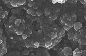
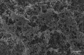
Left: Polypyrrole electrodeposited on conventional titanium. Right: Polypyrrole electrodeposited on multi-walled carbon nanotubes (Images: Webster Lab, Brown University)
Reporting their findings in the January 17, 2011 online issue of Nanotechnology ("Electrically controlled drug release from nanostructured polypyrrole coated on titanium"), Webster and his team demonstrate a 'proof of concept' to develop and evaluate on-demand delivery of penicillin/streptomycin (antibiotics used to treat grampositive and gram-negative bacteria) and dexamethasone (a glucocorticoid used clinically as an anti-inflammatory and immunosuppressive agent) in situ from polypyrrole. It shows that medications can be deployed on demand from the surface of bone implants to reduce the prevalence of both septic and aseptic complications.
This work is an extension of previous studies from Webster's nanomedicine lab demonstrating that these nanostructured materials sense and promote new bone growth ("Greater osteoblast functions on multiwalled carbon nanotubes grown from anodized nanotubular titanium for orthopedic applications"and "Multiwalled carbon nanotubes enhance electrochemical properties of titanium to determine in situ bone formation").
To create their polymer coating, the researchers first grew multi-walled carbon nanotubes (approx. 55nm in diameter) out of anodized nanotubular titanium using cobalt-catalyzed chemical vapor deposition. Polypyrrole monomers were either oxidized with the antibiotics or the dexamethasone before electrochemical polymerization of the polypyrrole around these nanotubes was carried out.
"Anionic drugs, bound electrostatically inside the polypyrrole thin film, were released in our present study by the application of a negative voltage," explains Webster. "For the first five cycles, we observed the anionic molecules to move in and out of the polypyrrole thin film due to continuous oxidation and reduction. The reduction peaks of penicillin/streptomycin release disappeared after 15 cyclic voltammetry cycles. For dexamethasone, the reduction peak was still observed after 25 cycles, but disappeared after 40 cycles."
The increase in the amount of drugs released after the electrical excitation was significant until 5 cycles. The cumulative release of penicillin/streptomycin and dexamethasone approached 80% of the drug and no further drug release was observed with further cycles.
Webster notes that, although they found that polypyrrole can be overoxidized and lose its electroactivity at higher potentials or when voltages are applied for longer periods, carbon nanotubes can maintain and prolong the electroactivity of polypyrrole due to their excellent conductivity properties.
He points out that polypyrrole can be doped not only with antibiotics and drugs like dexamethasone – allowing the preloading of drugs to obtain clinically controllable and predictable doses – but also various other biomolecules such as for instance growth factors, peptides, enzymes, antibodies, proteins, etc. to alter its biological, physical, chemical and electrical properties to design a controlled released system for numerous biomedical applications.
In addition, polypyrrole can be coated on electrodes or integrated with implantable chips to introduce an electrical signal into the biological environment.
"These preliminary results lay the foundation for developing intelligent orthopedic drug delivery technologies that can utilize a closed-loop sensing process with drug administration based on that sensing information," says Webster. "Both carbon nanotube-based sensors and controllable drug delivery systems could be an excellent way to improve the lifetime of orthopedic implants, allowing implants to kill bacteria, reduce the susceptibility of implants to prolonged inflammatory responses and ultimately increase bone formation."
By Michael Berger. Copyright 2011 Nanowerk

Monday, January 24, 2011

Professor Thomas Webster Named Scientist of the Week by Laboratory Equipment

Thomas WebsterEvery Thursday, Laboratory Equipment features a Scientist of the Week, chosen from the science industry’s latest headlines. This week’s scientist is Thomas Webster from Brown Univ. Webster and his team developed “liquid bone,” or a new kind of liquid that can be injected directly into broken bones and solidifies in seconds. Over time, new bone tissue will take its place, encouraged by natural growth factors embedded in the synthetic molecules of the material.
Q: Why did you decide to research and develop “liquid bone”? 
A: We received a lot of advice from medical doctors to explore materials other than titanium (or metals) for orthopedic applications. Basically, the clinical community was telling us to come up with something novel and more natural than what is being implanted today. The materials developed here, we call twin base linkers (or TBLs) originally come from DNA base pairs which are modified before use. Thus, we developed a material that comes from DNA. How natural is that!

We also got a lot of input from clinicians to develop materials which are injectable and thus would limit the amount of surgery required. Since the patient needs to heal from both the surgery and the new implant, the idea here was to limit the healing needed from the surgery by potentially using just a syringe to inject the material into the tissue defect. That way, healing time is minimized, and hopefully the healing process is accelerated to have quicker, healthier bone.

Q: Why do you feel this research is important to your field of study, and beyond?
A:
 I think, or rather hope, the material we developed will push the field to think outside of the box. We need to be really creative to create better implant materials. The fact that the hip implant has not changed that much since that first designed by Charnley in the 1960s disappoints me. So we are starting by throwing all conventional thinking aside to develop more natural nanostructured materials to be injected to heal tissues. Our tissues are naturally nanostructured, by the way.

Q: What was the most surprising thing you found in your research/process? 
A: The most surprising aspect of the material we developed is how quickly it solidifies into a material just as strong as bone. Usually, for injectable materials, you have to give up strength for fast solidification. By emulating DNA, we have created a material which can solidify within minutes inside the body and posses mechanical strength equivalent to bone. Of course, we can change the concentration of our material to have strengths less than bone and closer to other tissues, like cartilage.

Q: What is the take home message of your research and results? 
A: The bottom line from our approach and material is to look at the body to develop more natural materials and personalize such materials for the patient. For example, we can easily tailor the ability of these materials to grown bone to suite why a particular patient needs an implant. Consider an elderly female who broke her hip due a fall and that she has osteoporotic bone. Right now, that patient would essentially get the same implant as a teenager who was in a car accident. But our material could be modified for the elderly female to promote bone growth more than usual since her bones would be comprised in terms of their ability to grow bone. In a sense, we are creating materials for the future of personalized medicine. That can not be done with today's titanium.

Q: What is next for you and your research?
A: 
We need to do a lot more animal testing and then eventually human clinical trials. But we have a great team, with Dr. Hicham Fenniri from the Univ. of Alberta and our industry colleagues to help.

Compiled by Michelle Longo

Tuesday, January 18, 2011

Magnetically controlled pills could boost body’s absorption of drugs

Many drugs can only be absorbed in very specific parts of the intestine. In a new paper, Brown University scientists describe a new system that can safely hold a magnetic gelatin capsule in place anywhere in the gastrointestinal tract of a rat. In humans, the system could improve drug delivery and pharmacological research.
A place for everything
A tiny magnet inside a gelatin capsule allow researchers
to hold medicine at an exact place in the intestine
where it is best absorbed.
Credit: Mathiowitz Lab/Brown University 

PROVIDENCE, R.I.  — Do you want that in a pill or a shot?
Most patients never have that choice. The problem with administering many medications orally is that a pill often will not dissolve at exactly the right site in the gastrointestinal tract where the medicine can be absorbed into the bloodstream. A new magnetic pill system developed by Brown University researchers could solve the problem by safely holding a pill in place in the intestine wherever it needs to be.
The scientists describe the harmless operation of their magnetic pill system in rats online the week of Jan. 17 in theProceedings of the National Academy of Sciences. Applied to people in the future, said senior author Edith Mathiowitz, the technology could provide a new way to deliver many drugs to patients, including those with cancer or diabetes. It could also act as a powerful research tool to help scientists understand exactly where in the intestine different drugs are best absorbed.

Hold it right there!
As a magnet moves closer and farther from a small magnetic pill in a rat’s intestine, it keeps track of the force between it and the pill. The technology can be used to safely hold a pill in the right place to maximize absorption of the medicine it carries.
 Credit: Mathiowitz Lab/Brown University


“With this technology you can now tell where the pill is placed, take some blood samples and know exactly if the pill being in this region really enhances the bioavailability of the medicine in the body,” said Mathiowitz, professor of medical science in Brown’s Department of Molecular Pharmacology, Physiology, and Biotechnology. “It’s a completely new way to design a drug delivery system.”
The two main components of the system are conventional-looking gelatin capsules that contain a tiny magnet, and an external magnet that can precisely sense the force between it and the pill and vary that force, as needed, to hold the pill in place. The external magnet can sense the pill’s position, but because the pill is opaque to x-rays, the researchers were also able to see the pill in the rat’s bodies during their studies.
Safety first
The system is not the first attempt to guide pills magnetically, but it is the first one in which scientists can control the forces on a pill so that it’s safe to use in the body. They designed their system to sense the position of pills and hold them there with a minimum of force.
“The most important thing is to be able to monitor the forces that you exert on the pill in order to avoid damage to the surrounding tissue,” said Mathiowitz. “If you apply a little more than necessary force, your pill will be pulled to the external magnet, and this is a problem.”
To accomplish this, the team including lead author and former graduate student Bryan Laulicht took careful measurements and built an external magnet system with sophisticated computer control and feedback mechanisms.
“The greatest challenges were quantifying the required force range for maintaining a magnetic pill in the small intestines and constructing a device that could maintain intermagnetic forces within that range,” said Laulicht, who is now a postdoctoral scholar at MIT.
Even after holding a pill in place for 12 hours in the rats, the system applied a pressure on the intestinal wall that was less than 1/60th of what would be damaging.
The next step in the research is to begin delivering drugs using the system and testing their absorption, Mathiowitz and Laulicht said.
“Then it will move to larger animal models and ultimately into the clinic,” Laulicht said. “It is my hope that magnetic pill retention will be used to enable oral drug delivery solutions to previously unmet medical needs.”
In addition to Mathiowitz and Laulicht, authors on the paper include Brown researchers Nicholas Gidmark and Anubhav Tripathi. Brown University funded the research.

Thursday, January 13, 2011

Professor Thomas Webster edits biotech books


Thomas Webster, associate professor of engineering, has edited two recently published books. Nanotechnology Enabled In Situ Sensors for Monitoring Health is a compilation of recent research that uses the rapidly growing field of nanotechnology to design sensors to be used to diagnose and treat a variety of diseases. Biomaterials for Artificial Organs, which Webster edited with Michael Lysaght, a former Brown professor and director of the Center for Biomedical Engineering, reviews the latest synthetic materials often used as a substitute for organ transplantation

Monday, January 10, 2011

Brown Engineering Students and Alumni Organize "A Better World by Design" Conference

Brown engineering alumna Sharon Langevin '09 and current senior Tim Dingman '11 help organize the "A Better World by Design" annual conference with recent Brown graduate Steve Daniels. Dingman explains, “With students behind it, it’s more about entrepreneurship, about doing things. Actually, anyone can start their own organization and get things done. ABWxD is a very bottom-up process, which is the theme of the whole conference.” Their story was recently chronicled in a Providence Journal feature story.

Julia Steiny: Students want to do real things
01:00 AM EST on Sunday, January 9, 2011

The four were Sharon Langevin and Steve Daniels, from Brown University, and Tino Chow and Mike Eng from the Rhode Island School of Design (RISD). Together, they hatched a plan for what has since evolved into an annual conference called “A Better World by Design” (ABWxD). In the end, it changed their lives.In 2008, four hotshot college students had grown impatient with taking classes and digesting tons of information. They were itching to DO something. Somehow they wanted to get their hands dirty with changing the world, before finishing their fancy college educations.
The four wanted to hear from a cross-section of people who were solving real problems using social and environmental design ideas. With infectious idealism, they invited the coolest people they could find, from engineering, urban transportation, business, politics and so forth. With in-kind help from their respective institutions, but zero budget, they got engineers, architects and activists from all over the globe to agree to come.
To get the word out, they reached out to a local communications expert Andy Cutler. Cutler helped them think about generating free advertising, such as getting the speakers to promote the conference on their blogs, and designing communications with new media, new market strategies, new ways of getting things done.
Langevin says, “We had speakers lined up and we’d picked a weekend. But Andy got a lot more people involved, filling in workshops and panels with Brown and RISD professors.”
By the end of the summer, they had four more people working with them, but the task was monumental. Langevin shook her head and said, “We went into that weekend thinking that we would never do such a thing again. But we got such an amazing response, we came out of the weekend with: we HAVE to do this again next year.”
They did it. The confident idealists pulled off the sort of event that universities and corporations pay big bucks to professionals to pull off.
The first conference attracted 300 people. The next year it was 500. This past October it was 1,500.
I dropped in on a panel of professionals, each talking about work so mind-blowing it sounded like sci-fi. Aliza Peleg explained her company’s work designing fueling stations for electric cars that use robots to replace a spent car’s battery with a charged battery that the car owner rents.
Lisa Gansky, author of “The Mesh,” talked about moving from an ownership society to one in which more things are shared, like Zip cars, bike-share programs and apartment swaps.
Conference organizers made more effort to reach K-12 students, so Westerly middle-school kids presented their work on turning food grease into fuel.
The crowd buzzed with practical, entrepreneurial ideas. Invited speakers were found still talking together on the darkened stage where their presentation had long ago finished. Professionals from the Netherlands, Australia, India and the UK exchanged business cards and e-mails with each other and with the kids. Students from colleges as far away as Michigan found mentors to work with, summer internships and jobs, and new purpose.
Tim Dingman, one of this year’s organizers and a senior in engineering at Brown, explains, “With students behind it, it’s more about entrepreneurship, about doing things. Actually, anyone can start their own organization and get things done. ABWxD is a very bottom-up process, which is the theme of the whole conference.”
Sophia Yang from RISD, who is another current organizer, says, “When I got involved, I was transferring my interests from Furniture to Industrial Design. Furniture is an aesthetic and personal object. I wanted to do more than giving beautiful form. So the conference taught me how to use design to solve social problems. The conference itself was so powerful; I can’t say enough.”
As for Langevin, she finished her electrical engineering degree, but then went on to design a new Brown graduate program in International Development, which she is attending currently.
OK, so these are high-achieving students from top-league institutions. But if this group is impatient with abstract learning and not enough Doing, imagine how disempowered most urban students feel about their abilities to make the world a better place. Urban students, and others, are often trapped in communities that offer few opportunities and even fewer models of ordinary people using sheer will and personal charisma to tackle and solve a local problem.
Schools could be perfect places for students to connect to mentors and teachers who supervise them doing something concrete, reclaiming abandoned land or making other neighborhood improvements. As kids run into roadblocks, like advertising a conference without a budget, their mentors could help them find volunteers like Andy Cutler, who have lots of creative advice and expertise but no time to do the work for them.
It’s a different way of looking at education. Classes are not obsolete. In many ways even lecturing can be highly effective and efficient. But as the college kids found out, trying to get something done makes the learning in class much more relevant.
More importantly, though, urban kids need a far stronger sense of their own power to accomplish something they want. Given a chance, who knows what they might do.
Julia Steiny, a former member of the Providence School Board, consults on schools and government initiatives, such as Information Works!, Rhode Island’s school-accountability project. She can be reached at juliasteiny@gmail.com, or c/o EdWatch, The Providence Journal, 75 Fountain St., Providence, RI 02902.

Tuesday, January 4, 2011

Device Replicates Complex Bird Songs

A team of researchers, including Shreyas Mandre, have developed a simple rubber device that is able to replicate many different bird songs. The device may provide insight into how young birds learn songs from adults. Mandre is also working on a mathematical model to see if it is possible to identify some of the key principles in producing complex birdsong.


Simple rubber device mimics complex bird-song


The song is produced by blowing air through the device, which mimics a bird's vocal tract, the team explained.
The findings appear to challenge the idea that birds had to learn complicated neurological controls in order to produce distinctive calls.
The project's "holy grail" was replicating
the complex song of the zebra finch
The team plans to share its data with biologists to see if it sheds new light on how birds produce complex songs.
"I definitely did not think that I would be able to produce a whole bird song when we started," explained Aryesh Mukherjee, a member of the project team from Harvard University.
"We were just playing around and I probed the device in a certain way and it started playing a bird song - that was very exciting."
He added that the design of the device was very rudimentary: "It is made out of two pieces of rubber, which are stuck together but leaving a little area in the middle that forms the 'vocal tract'."
As well as the air source, the device is pressed together by a motor that replicates the action of a contracting muscle.
"In the terms of physics, the tract is just an elastic membrane of springs. If you tense it correctly, and probe it in a certain way, it starts vibrating," Mr Mukherjee told BBC News.
"Our project was to control the frequency of those vibrations."
The team were able to replicate a number of bird-songs, such as Bengalese finches and vireos, and were able to closely model the song of zebra finches.
"Making it sound like a zebra finch is the holy grail of the project," Mr Mukherjee said.
"We have been able to come pretty close to it, but we have been able to replicate other bird species much better."
He suggested that the song of the zebra finch was a little bit more complex, therefore it required a little fine tuning.
"But we are getting close," he added.
Good vibrations
The team's discovery was made during a project to learn more about the physical behaviour of vocal tracts.
"It was considered a very complicated process, and we tried to uncover some of the mysteries with physics.""We were working with neuroscientists who were trying to understand how a bird learns to sing.
Bird-song, a complex sound full of intricate patterns and rich harmonies, has been the subject of many studies. Neuroscientists, over the years, have provided insights into how young birds learn their songs from adult birds, requiring a series of complex neurological changes in order for them to control their voices.
But Mr Mukherjee said the project's results showed that it was possible to replicate bird-song without high degrees of control inputs.
"By just having one muscle (motor pressing the device) in the equation, you can get a lot of sounds," he explained.
The surprisingly simple design was able to replicate
complex birdsong of a range of species
"Translating that back into the idea of neurological control... it suggests that the control needed to produce seemingly complex songs is not as challenging as previously thought."
However, Mr Mukherjee said that whether this challenges current thinking on how birds produce their song was outside their area of expertise.
"We are in no position to make a claim about what this has to do with bio-physics or neurological control within birds. All we can say is what we have learned from our experiments, and share that information with biologists.
Another member of the team Shreyas Madre - now an assistant professor at Brown University, Rhode Island - is developing a mathematical model to see if it is possible to identify some of the key principles in producing complex birdsong.
The team plans to publish its findings in a paper in the near future.

Monday, January 3, 2011

Time Magazine Calls Artificial Ovary Developed by Brown University Researchers One of Top 10 Medical Breakthroughs

Time Magazine's Top 10 Medical Breakthroughs of 2010


9. Artificial Ovary

RALF HIRSCHBERGER / EPA / CORBIS
In more good news for those struggling with infertility, scientists reported success in creating an artificial ovary that could one day nurture immature human eggs outside the body. Researchers led by a team at Brown University managed to coax three primary ovary cells donated by patients into a 3-D structure resembling an ovary. In the lab, the cell types interacted with one another and functioned for all intents and purposes like a real ovary, even successfully maturing a human egg from its earliest stages in the follicle to a fully developed form.

Most immediately, the structure could help IVF technicians improve success rates. Currently when women donate eggs for a cycle of IVF, they provide a range of both mature and immature eggs; the less developed ones are less likely to be fertilized to become embryos. But by allowing technicians to mature these eggs in the lab, researchers might be able to help each IVF cycle become more efficient in leading to a pregnancy and eventual life birth. In addition, the artificial ovary could help women with ovarian disease, who are unable to produce mature eggs, take advantage of IVF to have children of their own.


Researchers build ‘artificial ovary’ to develop oocytes into mature human eggs

Researchers at Brown University and Women and Infants Hospital of Rhode Island have built an artificial human ovary that can grow oocytes into mature human eggs in the laboratory. That development, reported in the Journal of Assisted Reproduction and Genetics, could help preserve fertility for women facing chemotherapy or other treatments.
An artificial ovaryAn engineered honeycomb of cultured theca cells
(top row) envelopes spheres of granulosa cells (GC).
The bottom row shows the tissue after 48 hours (left)
and after five days.
Credit: Carson Lab / Brown University
PROVIDENCE, R.I. [Brown University] — Researchers at Brown University and Women and Infants Hospital have invented the first artificial human ovary, an advance that provides a potentially powerful new means for conducting fertility research and could also yield infertility treatments for cancer patients. The team has already used the lab-grown organ to mature human eggs.
“An ovary is composed of three main cell types, and this is the first time that anyone has created a 3-D tissue structure with triple cell line,” said Sandra Carson, professor of obstetrics and gynecology at the Warren Alpert Medical School of Brown University and director of the Division of Reproductive Endocrinology and Infertility at Women and Infants Hospital. Carson is a senior author of a recent article in the Journal of Assisted Reproduction and Genetics that describes the innovation.
Carson said that the ovary not only provides a living laboratory for investigating fundamental questions about how healthy ovaries work, but also can act as a testbed for seeing how problems, such as exposure to toxins or other chemicals, can disrupt egg maturation and health.
Clinically, the artificial ovary could play a role in preserving the fertility of women facing cancer treatment in the future, said Stephan Krotz, a Houston fertility doctor who is the paper’s lead author and a former fellow in Carson’s lab. Immature eggs could be salvaged and frozen before the onset of chemotherapy or radiation, he said, and then matured outside the patient in the artificial ovary.
Building an ovary
What makes the artificial ovary a functional tissue, rather than just a cell culture, is that it brings all three ovarian cell types into a 3-D arrangement similar to a real ovary in the body. The means for making such compositions of cells was invented in the lab of Jeffrey Morgan, associate professor of medical science and engineering, who is a co-author of the paper published online Aug. 25. His so-called 3D Petri dishes are made of a moldable agarose gel that provides a nurturing template to encourage cells to assemble into specific shapes.
To create the ovary, the researchers formed honeycombs of theca cells, one of two key types in the ovary, donated by reproductive-age (25-46) patients at the hospital. After the theca cells grew into the honeycomb shape, spherical clumps of donated granulosa cells were inserted into the holes of the honeycomb together with human egg cells, known as oocytes. In a couple days the theca cells enveloped the granulosa and eggs, mimicking a real ovary.
The big test, however, was whether the structure could function like an ovary — namely to mature eggs. In experiments the structure was able to nurture eggs from the “early antral follicle” stage to full maturity.
“[This] represents the first success in using 3-D tissue engineering principles for in vitro oocyte maturation,” the researchers wrote in the journal article.
Carson said her goal was never to invent an artificial organ, per se, but merely to create a research environment in which she could study how theca and granulosa cells and oocytes interact. When she learned of Morgan’s 3-D Petri dishes, they began to collaborate on creating an organ. Morgan said this is the first fully functional tissue to be made using the method.
To help fund the work, Morgan and Carson applied for and won a Collaborative Research Award from the Rhode Island Science and Technology Advisory Council (STAC). STAC grants encourage research with commercial potential. Morgan has recently founded a local Rhode Island startup, MicroTissues Inc. The company will begin selling these micro-mold tools in about a month to researchers looking to engineer 3-D tissues. Other funding came from Women and Infants Hospital.
With what appears to be a fully functional artificial organ, Carson and Morgan continue to collaborate and are now embarking on the studies she dreamed it would make possible. She’s reluctant to predict what they’ll turn up, however.
“This is really very, very new,” she said.
The paper’s other authors are Jared Robins, Toni-Marie Ferruccio, Richard Moore and Margaret Steinhoff, all of Brown University.

Robot Reveals Unusual Fish’s Movement

A team of researchers, including Oscar Curet, postdoctoral research associate in engineering, have developed a robot that mimics the movement of the black ghost knifefish. Called the GhostBot, the machine reveals how the finless fish maneuvers through the murky waters of the Amazon Basin. “Animals usually never behave the way you need them to do while studying their movements. With robotics that mimic animal movement, you do something over and over again without hurting the animal,” Curet says.


Black Ghost Knifefish Robot Unmasks Movement Secrets

Borrowing biological designs from the black ghost knifefish, engineers have built a swimming robot that reveals how the animal’s trick of vertical movement works.
Called GhostBot, the robot copies the real fish’s undulating, ribbon-like ventral fin to propel itself through the water. New high-speed experiments show how, when waves travel along the robot’s ribbon from head to tail and meet in the middle, mushroom-cloud-like jets can push it upward.
“These fish are extremely maneuverable, and we knew how they move forward and backward with their fins,” said bioengineer Malcolm MacIver of Northwestern University, who led GhostBot’s design. “What we didn’t know was how they move vertically.”
The black ghost knifefish lives in the rivers of the Amazon Basin, using a self-generating electric field to see through the murky waters. It doesn’t have traditional fins like a bony fish, nor does it sway its body like an eel to move around. Instead, it stays rigid and uses a ribbon-like fin along its belly to navigate through a maze of downed trees, stones and other underwater obstructions with extreme precision.
Emulating such maneuverability in robotic submersibles would create countless opportunities for new or more robust underwater research, says MacIver, co-author of a study detailing the vertical movement mechanics in an upcoming issue of the Journal of the Royal Society Interface. An efficient, submersible hovering robot, for example, could constantly monitor the health of a coral reef without crashing into it (most researchers hire costly divers to do the work).
“I don’t agree that nature always has the best designs, but this is a place where it’s way ahead of human technology,” MacIver said.
When a knifefish moves upward in water, it rolls two opposing waves down its fin that meet in the middle. How that action generates upward thrust — which the dense fish needs, otherwise it’d sink — was a puzzle until the research team, including mechanical engineer Oscar Curet (now at Brown University), decided to build a copycat robot.
“Animals usually never behave the way you need them to do while studying their movements,” Curet said. “With robotics that mimic animal movement, you do something over and over again without hurting the animal.”
To reveal how the black ghost and other knifefish move vertically, the scientists worked with a company called Kinea Design to build GhostBot. It took them about seven months and $200,000 to complete a finished version.
They put the forearm-sized device into a special tank able to reveal the fluid mechanics near the fin. It flowed water laced with shiny particles over the swimming robot, shined a laser-beam plane onto the ribbon fin and took footage with high-speed cameras. Computer algorithms then processed the images to map particle velocities.
“While swimming upward, two opposing jets of water meet in the middle and collide. The merged jets deflect downward and push up on the fish,” Curet said. The animal can modulate the jets to move in diagonal directions as well, he says.
“This kind of ribbon fin is something worth paying attention to, because it has independently evolved around the world many times,” said Noah Cowan, a biologist and mechanical engineer at Johns Hopkins University who wasn’t involved in the study. “It’s a very robust design by nature, and these particular animals can move with very little body bending. It may be good for moving a rigid submersible robot.”
A major robotics company interested in developing autonomous submersibles is already discussing how they can use the design, MacIver says. Attaching ribbon-like mechanical fins to submarines people fit into, however, is another matter.
“It sounds fantastic for autonomous submersibles, but we may never use it. The design seems too fragile to sit on the ocean floor, which is what customers sometimes do with our submarines,” said Patrick Lahey, president of Triton Sumarines. “For safety reasons, we have to adhere to tried and tested technologies.”
In addition to GhostBot’s unusual means of swimming, MacIver says it’s also packed with sensors emulating the real animal’s electrosensing abilities.
“They basically use their body as a big eye,” he said. “We want to connect the robot’s swimming to its sensor network and show it can autonomously find an object we want, then hover to say, ‘here it is.’”
Videos: 1) High-speed footage of a swimming black ghost knifefish, the submersible GhostBot, and fluid mechanics experiments performed on the device. Credit: Journal of the Royal Society Interface. 2) A black ghost knifefish swims in strange directions to find and eat food. Credit: YouTube.
Image: Fluid mechanics models showing how the black ghost knifefish’s vertical jets form and push upward on its body. Credit: Journal of the Royal Society Interface.